AI-Assisted ECG Interpretation for Subclinical Cardiac Disease
Deep learning finds cardiac disease hidden in ECGs that doctors cannot see.
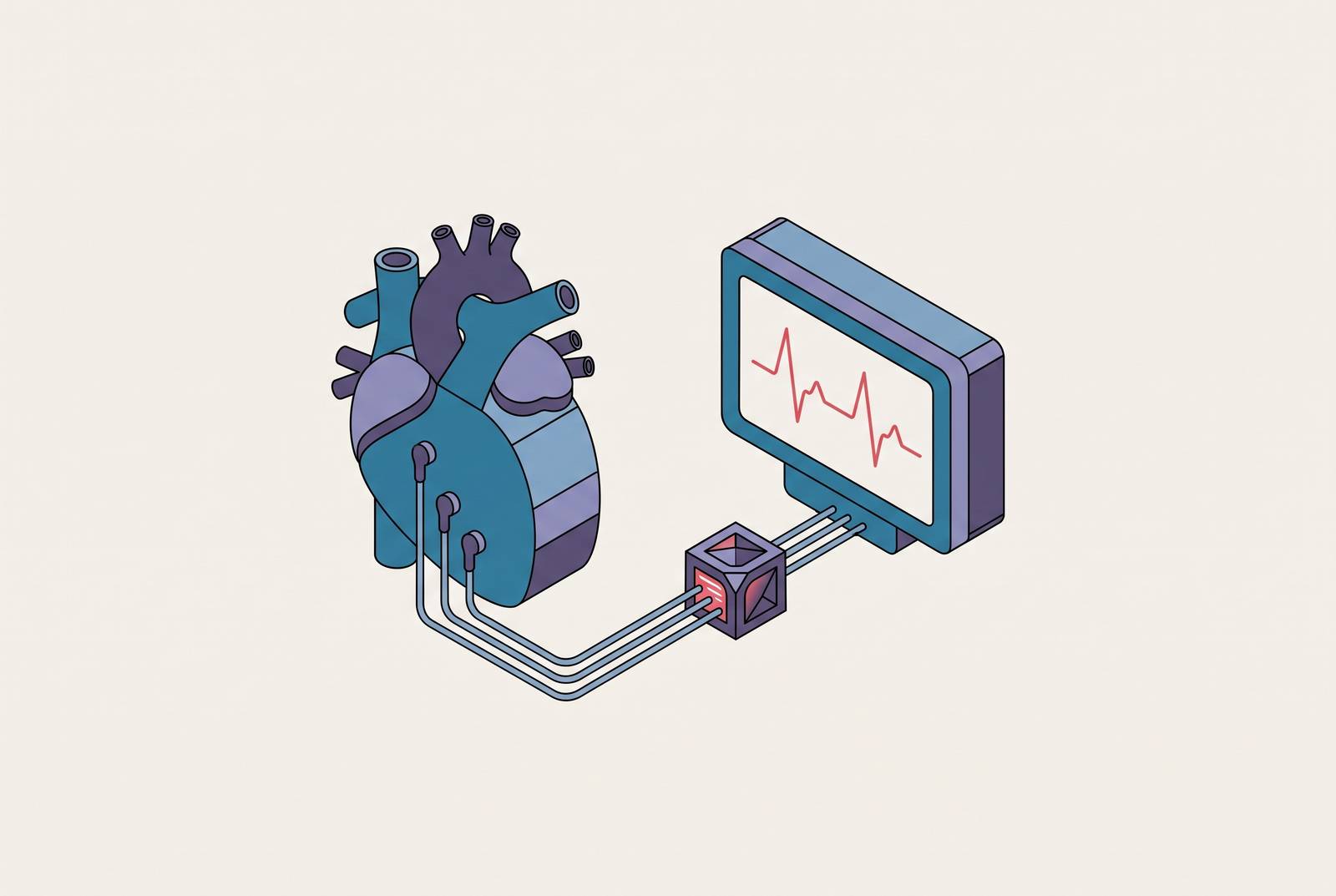
The electrocardiogram is the most widely used cardiac test in medicine. It is cheap, fast, non-invasive, and available in nearly every clinical setting on earth, from a rural clinic to a tertiary academic hospital. That ubiquity matters because cardiovascular disease kills about 17.9 million people a year, and the ECG is often the first, and sometimes the only, cardiac test a patient ever gets. But the tool's reach has never matched its resolution. A standard 12-lead ECG produces a waveform with dozens of measurable features, and for over a century, the job of reading it has fallen to a human eye trained to spot a fairly narrow set of visual patterns, including ST elevation, a widened ventricular depolarization complex, and a prolonged QT.
That process is subjective in ways the field has always known about but rarely reckoned with directly. Inter-observer and intra-observer variability are well documented; the same tracing read by two cardiologists, or by the same cardiologist on two different days, can yield different conclusions. Fatigue, training level, and clinical context all shift the read. None of this is a knock on clinicians. It's a structural limit. Human pattern recognition, however well trained, was never built to detect the kind of high-dimensional, sub-visual signal that reflects microstructural remodeling in heart muscle or electrical tissue years before it becomes symptomatic.
That's why sensitivity for subclinical cardiomyopathies, incipient left ventricular dysfunction, and future atrial fibrillation stays modest under conventional interpretation. The abnormality is often already encoded in the waveform. It's just not visible to the kind of pattern-matching a human reader does. The ECG hasn't been the bottleneck. The eye reading it has.
How deep learning extracts what human readers cannot see
Rule-based ECG software, the kind that has flagged intervals, amplitudes, and axis deviations for decades, works from a fixed list of features someone decided mattered. Deep learning does something categorically different. Trained on large sets of annotated ECGs, these models find statistical patterns that were never named as diagnostic criteria in the first place, patterns that exist somewhere in the relationship between thousands of waveform data points rather than in any single measurable interval.
Convolutional neural networks (CNNs) do most of the heavy lifting here. They were built for image and signal pattern recognition, and applied to ECGs, they've shown strong performance in automated arrhythmia classification and in flagging subclinical structural disease, including reduced LV function, hypertrophic cardiomyopathy, and cardiac amyloidosis. Long short-term memory (LSTM) networks bring something different to the table: they're built to model sequences over time, which suits the temporal, beat-to-beat structure of an ECG signal. Hybrid CNN-LSTM architectures try to get both, spatial pattern extraction and temporal sequence modeling, in a single pipeline.
Signal denoising and other plumbing make the rest of this possible. Signal denoising pipelines strip out motion artifact and baseline wander before a model ever sees the data. Synthetic data generation addresses the class imbalance baked into any dataset where the disease being hunted is, definitionally, rare. And explainable AI (XAI) techniques matter for a different reason: without some way to surface which part of the waveform actually drove a given prediction, a model's output is a black box a clinician has no principled reason to trust or distrust. XAI doesn't just help adoption. It's how anyone finds out whether a model has learned physiology or learned a shortcut.
Detecting silent and paroxysmal atrial fibrillation before a stroke occurs
Atrial fibrillation raises stroke risk by as much as fivefold, and in patients whose AF goes untreated, stroke incidence has been reported in the range of 7.7 to 30.8 (per relevant cohort measure). The trouble with AF has always been catching it before the stroke, particularly in its paroxysmal form, since diagnosing it once it's on the tracing has never been the issue. It's catching it before the stroke, particularly in its paroxysmal form, when the arrhythmia comes and goes and a routine ECG has a good chance of catching the patient in normal sinus rhythm.
AI-ECG reframes the entire task. Instead of waiting to detect overt AF on a tracing taken during an episode, models now try to infer AF risk, or predict future onset, from a sinus-rhythm ECG that looks entirely unremarkable to a human reader. The signal being read is some subtler electrical fingerprint, not the arrhythmia itself. It's some subtler electrical fingerprint, sometimes derived from heart rate variability, sometimes pulled from the raw waveform, that correlates with a heart that will develop AF at some point down the line. Prediction horizons in this line of research have ranged from minutes to years out, which matters practically: it turns AF risk stratification into something that can guide who gets a monitoring patch or an implantable loop recorder, rather than requiring blanket screening of every patient who walks through the door.
The VITAL-AF trial tested this idea in primary care, screening patients 65 and older with a handheld single-lead ECG. In a test set of 4,221 individuals, an AI model trained on single-lead ECG data achieved two-year AF discrimination comparable to models trained on the full, richer VITAL-AF dataset. A cheap, single-lead handheld device, the kind that fits in a primary care visit without any special equipment, can rival the performance of models built on far more data-rich inputs. A screening tool that stays confined to academic cardiology and one that scales into a waiting room depend on that.
Identifying reduced left ventricular ejection fraction from a routine ECG
Reduced LV ejection fraction is frequently silent until it isn't. Patients can walk around with meaningfully impaired systolic function and no symptoms at all, until the disease has progressed far enough to become heart failure with a hospitalization attached. That makes it a near-ideal target for opportunistic screening, especially because, unlike many conditions AI-ECG is chasing, disease-modifying therapy already exists once reduced LVEF is caught. The bottleneck is detection. It's detection.
The central claim in this research area is a strange one to sit with: the ECG can look completely normal to a trained human reader, showing none of the conventional red flags for LV dysfunction, and still contain enough signal for a model to flag it. Mayo Clinic's deep learning model, one of the most cited in this space, predicts reduced LVEF (at or below 35%) in the general population with an AUC of 0.93. That's a strong number by any diagnostic standard: an AUC that high buys a clinician a tool that can rule in or rule out a serious, treatable condition using a test that already gets ordered for other reasons, with no added cost or radiation or invasive step.
What makes that number more than an academic curiosity is what happened after it: multicenter external validation of ECG-AI software as a medical device for LVEF detection, run across four geographically diverse sites in one country. sites, using real-world patients who happened to have both an ECG and a transthoracic echocardiogram within 30 days of each other in the normal course of care. That's the exact kind of pragmatic, multi-site validation regulatory pathways like FDA submission are built around, and it's a meaningfully higher bar than a single-center retrospective study. Low-LVEF detection is, at this point, the AI-ECG use case with the most mature evidence base behind it.
Structural heart diseases where AI-ECG evidence is earlier or incomplete
The AI-ECG literature addresses a range of conditions where the technology may serve as a pre-echocardiographic triage tool, spanning a range of structural conditions, including reduced LVEF, hypertrophic cardiomyopathy, cardiac amyloidosis, and composite models that try to flag several conditions from one tracing. Not all of these sit at the same point on the evidence curve, and conflating them does a disservice to how differently each has been validated.
A distinction that matters more than it might sound like on first read is whether AI-ECG serves as a safety-net or a gatekeeper. In a safety-net role, a positive AI-ECG result adds a trigger for confirmatory echocardiography, essentially widening the net of who gets a closer look. In a gatekeeper role, a negative or low-risk AI-ECG result gets used to justify deferring or skipping echocardiography altogether in select low-risk settings. Those two uses are not equivalent in what they risk. A safety-net tool that over-triggers wastes an echocardiogram. A gatekeeper tool with an unacceptable false-negative rate lets real disease walk out the door undetected, which is a categorically more dangerous failure mode and requires a much higher evidentiary bar before deployment.
Judged against that bar, low-LVEF detection is furthest along, backed by pragmatic randomized trial data and multicenter external validation. Valvular disease and composite structural heart disease models show promise for enriching referral pathways, catching patients who'd benefit from a cardiology workup they weren't otherwise going to get. HCM, cardiac amyloidosis, and pulmonary hypertension sit earlier on that curve, either with an incomplete evidence pathway or validation that hasn't yet cleared the bar the LVEF models have.
Cardiac amyloidosis is the clearest illustration of both the promise and the limits still baked into this technology. Post-development validation of the Mayo Clinic AI-ECG model for amyloidosis has shown strong aggregate performance, and results have generally held up across key subgroups in aggregate. But performance dropped in patients with left bundle branch block, in those with LV hypertrophy, and in ethnically diverse populations specifically. That's the exact subgroup-specificity problem that keeps a strong average AUC from translating cleanly into a tool that's safe to deploy across every patient who walks in the door. It's the exact subgroup-specificity problem that keeps a strong average AUC from translating cleanly into a tool that's safe to deploy across every patient who walks in the door.
Evidence gaps: generalizability, bias, and the validation gap
Generalizability produces nearly every other limitation in this field, as the evidence below shows. Most published AI-ECG models were built and trained on data from a single large academic medical center, or from a national population that's demographically fairly uniform. Performance that looks excellent in that setting doesn't reliably survive the trip to a different hospital system, a different country, or a patient population with a different mix of age, race, and comorbidity.
The AF prediction literature gives a concrete number to hang that concern on: across multiple studies using the same underlying model, discrimination for AF prediction ranged from an AUC of 83.8 down to 73.2, reflecting meaningfully different real-world performance across validation settings. Same model, same clinical question, meaningfully different real-world performance. That's not a rounding error; it's the difference between a tool that's clinically useful and one that isn't, depending on where a patient happens to live.
The amyloidosis model's documented weak spots, including certain comorbid conditions and ethnically diverse populations, sit at the intersection of two distinct failure types: one is a structural confound (a conduction abnormality that changes the waveform in ways that can mask or mimic the target pattern), and the other is a demographic gap tied directly to who was in the training data. Multiple reviews trace both problems back to the same root cause: training datasets that underrepresent certain demographic groups, combined with insufficient validation of these models in real-world, non-academic clinical settings before they get near a patient.
Requirements for Clinical Adoption: Workflow Integration, Decision Support Design, and the Clinical Evidence Bar
None of this technology does anything for a patient sitting in a spreadsheet or a validation paper. Getting an AI-ECG model from a strong AUC to a tool that changes practice requires it to sit inside a workflow a clinician already uses, without adding friction that makes them stop trusting it or stop using it. That means the output has to show up where an ECG already gets read, as part of the existing workflow rather than a separate application requiring a separate login, and it has to communicate a risk signal in language a primary care physician or a nurse practitioner can act on without a cardiology fellowship.
Decision support design carries real weight here, and the safety-net versus gatekeeper distinction from the structural heart disease evidence functions as a real constraint on deployment, not just an academic framing device. It's a design decision with direct consequences for how a false negative or false positive gets handled downstream. A tool built to flag more patients for a confirmatory echo can tolerate a certain amount of noise. A tool built to justify skipping that echo cannot, and deploying the second kind with evidence suited only to the first is where oversight bodies and health systems alike should draw a hard line.
The clinical evidence bar, ultimately, is the whole story here. An AUC from a single academic center's retrospective dataset is a promising first finding for changing how care gets delivered. What the field needs more of, and what the LVEF work has started to provide, is multicenter external validation run across geographically diverse sites using real-world patients, representing a meaningfully higher bar than single-center retrospective studies, before any of this moves from research pipeline to routine practice. The ECG has always been the most democratized test in cardiology. What AI adds is a measurable gain in detection, and the validation must be done right, not rushed, given stakes as large as the promise the technology carries.
Sources
- Advances in the Interpretation of the Electrocardiogram by Artificial Intelligence | MDPI
- Advances in the Interpretation of the Electrocardiogram by Artificial Intelligence
- Systematic Review of Artificial Intelligence and Electrocardiography for Cardiovascular Disease Diagnosis | MDPI
- Investigating the Efficacy of AI-Powered Innovations in ECG Analysis and Continuous Heart Monitoring: A Comprehensive Narrative Review
- Systematic Review of Artificial Intelligence and Electrocardiography for Cardiovascular Disease Diagnosis
- AI-enhanced electrocardiography as a digital biomarker platform in cardiovascular medicine: clinical applications, validation gaps and future implementation pathways
- A Vision Transformer for ECG-Based Detection of Left Ventricular Systolic Dysfunction Across Multiple Clinical Sites
- Artificial intelligence in arrhythmia risk prediction: connecting undetectable vulnerabilities and proactive care