MRI vs Mammography for Breast Cancer Screening in High-Risk Women
MRI detects twice as many cancers in high-risk women despite more false alarms.
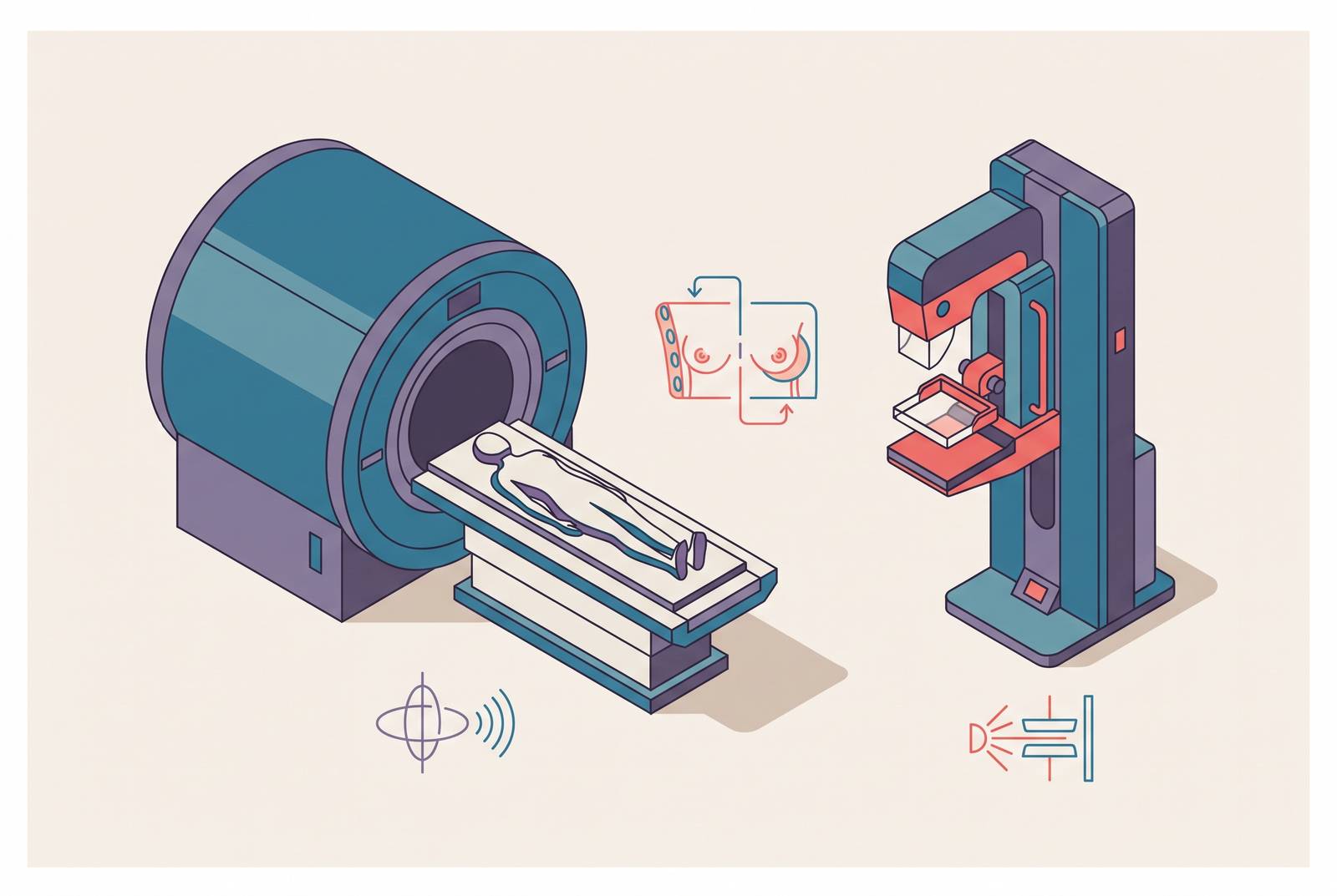
Breast cancer screening in women at high lifetime risk runs into a structural problem: mammography, the tool built for the general population, performs worst in exactly the biology that defines this group. Dense tissue and fast-growing tumors undercut the X-ray mechanism at its core, and that is why MRI, even though it catches more false positives, has become the recommended first tool for high-risk screening, not just a supplement bolted onto mammography.
Why mammography struggles in high-risk women
Mammography works by passing X-rays through breast tissue and reading the differences in how tissue types absorb them. A tumor shows up because it attenuates X-rays differently than the fat and glandular tissue around it. But when the surrounding tissue stops being a helpful, low-density backdrop, that mechanism runs into trouble. Dense fibroglandular tissue absorbs X-rays at rates close to those of tumors themselves, and the result is that mammography sensitivity can drop by 30 to 50 percent in dense breasts. The tissue doesn't just make the scan harder to read; dense tissue is also, independently, a known risk factor for developing cancer. So the same women who carry more fibroglandular tissue, and therefore more risk, are also the women in whom a mammogram is least equipped to find a tumor if one is there.
That's a problem of physics, not of effort or technique on the radiologist's part. The tumor biology seen in high-risk populations, particularly in women carrying BRCA1 mutations, layers a second mismatch onto this one. These tumors tend to grow fast. A lesion that wasn't there, or wasn't visible, at last year's mammogram can become clinically significant well before the next annual scan comes around. No amount of image clarity solves a timing problem. If a screening interval is built around slower-growing, average-risk disease, it isn't matched to how quickly these cancers can develop and spread.
Younger women make up a large share of the population classified as high-risk, through family history, genetic testing, or prior radiation exposure, and because younger breast tissue tends to be denser and their tumors tend to grow faster, both problems land on the same patients at the same time, compounding each other.
MRI gets around the density issue by using an entirely different physical signal. Rather than reading differences in X-ray attenuation, breast MRI tracks how tissue enhances after a contrast agent is injected, based on blood flow and vascular permeability, properties that have nothing to do with tissue density. A tumor's blood supply tends to behave differently from surrounding tissue regardless of how dense that tissue is on a mammogram. Mammography relies on tissue contrast, a mechanism density itself undermines. MRI simply doesn't have it.
How large the sensitivity gap is
The sensitivity difference between the two modalities in high-risk women is a gap wide enough to change how early a cancer gets caught, holding up across several different study designs. The Dutch MRISC trial, one of the most cited prospective studies in this field, found MRI sensitivity for invasive cancer running at more than twice that of mammography, while mammography held a modest specificity edge over MRI. In a population already carrying an elevated baseline risk of cancer, missing a cancer costs far more than a false alarm does.
A meta-analysis pooling 11 prospective comparative studies found the same pattern at scale. When annual contrast-enhanced MRI was added to mammography, combined sensitivity rose above 90 percent, more than double what mammography achieved on its own. You can trace it across a decade-plus of prospective research in high-risk cohorts screened on different continents.
What that sensitivity gap buys, clinically, is stage. A cancer caught while still small and node-negative carries a far better prognosis than one found after it has spread to lymph nodes or beyond, and that isn't a statistical artifact of earlier labeling. It reflects the actual biological window screening is meant to exploit, catching disease before it progresses to a point where treatment options narrow and outcomes worsen. Protocol documentation for the ongoing trial registered as NCT03220893 reports that serial MRI screening over several years markedly reduced the rate of late-stage disease at presentation compared with controls. Finding cancer earlier is the entire mechanism by which screening is supposed to prevent death, and a sensitivity gap this large, sustained over years of screening, is what makes that mechanism function in a high-risk population.
The real cost of MRI's lower specificity
MRI's weaker specificity is a legitimate concern, not a footnote to wave away. In absolute terms, MRI triggers more false positives than mammography does. Some studies report recall rates as high as 26 percent, and when programs run supplemental MRI alongside mammography, they report meaningfully higher false-positive counts per screening round than mammography alone produces. Any honest comparison has to start by admitting that.
What changes the calculation is what happens once a biopsy gets ordered. Research comparing biopsy outcomes across both modalities has found that the positive predictive value of biopsy, the share of biopsies that actually turn up cancer, runs similar for MRI and for mammography. A biopsy triggered by an MRI finding is roughly as likely to find cancer as one triggered by a mammography finding. The two tests generate different kinds of false alarms too. MRI false positives tend to be high-risk proliferative lesions with atypia, the kind of finding a pathologist takes seriously even when it isn't cancer. Mammography false positives skew toward low-risk, non-proliferative changes. MRI's false alarms are, more often than not, findings with some clinical weight behind them.
Specificity also behaves differently depending on who's being tested. The same specificity figure does less damage in a population where the prior probability of cancer is already high than it does in a general screening population, because a positive result in a high-prior population is simply more likely to reflect real disease. That's basic test-performance math: a given false-positive rate carries a smaller relative cost in a high-risk cohort than the same rate would carry if it screened average-risk women.
None of that erases the human cost of a false alarm. Unneeded callbacks, unneeded biopsies, and the anxiety that comes with both are burdens on patients, and the honest version of this argument has to carry that weight. Overdiagnosis deserves the same treatment. A more sensitive test can also catch small, slow-growing tumors that would never have caused harm in a woman's lifetime, and that risk applies to MRI just as it would apply to any screening tool more sensitive than what came before it. Trial background for the MRIB study raises this concern directly. The honest accounting still tilts toward MRI in this specific population, but it tilts there with real costs attached, not for free.
Evidence and the critical gap
The case for MRI rests on detection and staging data that are large, repeated, and consistent across trial designs. What that evidence does not yet include is a randomized trial that tracks women screened with mammography alone against women screened with MRI plus mammography all the way to mortality. Susan G. Komen states this gap explicitly, and it's the strongest objection a careful critic can raise against the current standard of care.
The logic connecting earlier detection to longer survival is well established in cancer biology generally, and it holds up across the broader mammography literature: finding cancer at an earlier stage should, as a matter of tumor growth and metastatic spread, improve survival odds. But that logic has not been closed with trial data measuring mortality directly in this specific comparison, MRI against mammography, in high-risk women. The trials driving the current evidence base, MRISC, MARIBS, and the 12-center randomized trial, are rigorous studies with prospective designs, and they are strong on detection outcomes. Detection is still a surrogate endpoint, standing in for the outcome that actually matters. Critics can point to instances across medicine where a surrogate endpoint looked convincing and the mortality data later told a more complicated story, and that history deserves to be taken seriously here too.
A further concern sits outside the detection-versus-survival debate entirely: gadolinium, the contrast agent used in breast MRI, accumulates in the body with repeated exposure over years of annual screening. Trials designed to measure detection and staging outcomes were not built to track that accumulation or its long-term effects, so this risk remains largely unaddressed by the existing evidence base.
None of that undercuts the case for MRI. It's precisely the kind of evidence, consistent, large in magnitude, and biologically coherent even without a closed mortality loop, on which clinical guidelines are routinely built. Medicine rarely waits for a perfectly closed causal chain before acting on a pattern this strong, and the guidelines discussed below reflect that judgment rather than a claim that every question has been answered.
The benefit of adding mammography to MRI by age and mutation status
The real clinical question is which women benefit from adding mammography on top of MRI, and which women see no meaningful gain from the combination. The answer depends heavily on age and mutation status, and it refines the case for MRI.
A large prospective cohort of high-risk women found that adding mammography to MRI in BRCA mutation carriers aged 30 to 39 added essentially no extra sensitivity. MRI alone performed about as well as the combination in that age group. Once women pass 50, the picture shifts: breast density typically declines with age, mammography's X-ray contrast improves as a direct result, and combining MRI with mammography produced a statistically significant sensitivity increase over MRI alone. The two tools become genuinely complementary once density is no longer the dominant obstacle standing between the X-ray beam and the tumor.
A 2025 systematic review and meta-analysis backed up the younger-patient finding: combined MRI and mammography offered no significant sensitivity gain over MRI alone in BRCA1 mutation carriers or in women under 40. That finding reopens a fair question: whether mammography's radiation exposure and weaker specificity are worth imposing on the youngest, highest-risk patients when MRI alone appears to catch what the combination catches.
A retrospective cohort from the Mercy Breast Clinic in Auckland, New Zealand, covering May 2022 through September 2023 and including a large group of women classified as high-risk by Tyrer-Cuzick v8 score, found that MRI and ultrasound detected invasive cancers that were occult on every mammographic view, concentrated mainly in women with density D breasts, the densest classification. The same cohort showed MRI and ultrasound outperforming mammography at mapping disease extent, including multifocal and multicentric disease. That data comes from a preprint that has not yet completed peer review and should be read with that caveat attached.
Taken together, these findings don't argue for retreating to mammography-first protocols. They argue for applying MRI-led screening with some precision: MRI as the backbone for younger BRCA1 carriers where mammography adds little, and MRI plus mammography for older high-risk women where the combination earns its keep.
What current guidelines recommend
Clinical guidelines have caught up to this evidence and now treat MRI as the lead tool for high-risk screening. The 2025 ACR Appropriateness Criteria recommend starting breast MRI as early as age 25 to 30 in high-risk patients, with mammography or digital breast tomosynthesis layered in starting anywhere from age 25 to 40 depending on the type of risk a patient carries. MRI comes first in that sequence. Mammography supports it.
In April 2026, the American Society of Breast Surgeons took a related but separately framed position: it recommends annual 3D screening mammography for higher-than-average-risk women, with enhanced surveillance through annual breast MRI considered when indicated. The two statements don't contradict each other so much as reflect how different professional bodies weigh sequencing and emphasis, while agreeing that both tools belong in the regimen for this population.
Current ACR recommendations also name contrast-enhanced mammography as an option for women who cannot undergo MRI, whether because of contraindications like certain implanted devices or because of access and availability constraints. That inclusion matters beyond its practical use: it signals that the actual goal is MRI-equivalent sensitivity, not loyalty to MRI as a specific technology, and that the field is actively building alternatives for settings where MRI isn't a realistic option.
When women with extremely dense breasts had negative mammograms, supplemental MRI produced a significantly lower rate of interval breast cancers (cancers diagnosed between scheduled screenings) than mammography alone. It stands as the most-cited randomized evidence behind dense-breast screening recommendations, and it fills in, for the dense-breast subpopulation specifically, some of the randomized rigor that the broader MRI-versus-mammography comparison still lacks.
Guidelines built this way, MRI starting early, mammography calibrated to age and risk type, alternatives named for women who can't access MRI, reflect a field that has read the detection evidence carefully and adjusted the standard of care accordingly, even while acknowledging, as the evidence section above does, that the mortality data closing the final loop is still being gathered.
Sources
- ACR Appropriateness Criteria® Female Breast Cancer Screening: 2025 Update - Journal of the American College of Radiology
- Improving Breast Cancer Detection in Higher Risk Women: A Multimodality Imaging Evaluation in a Private Screening Clinic
- The added value of mammography in different age-groups of women with and without BRCA mutation screened with breast MRI
- MRI versus mammography plus ultrasound in women at intermediate breast cancer risk: study design and protocol of the MRIB multicentre randomized controlled trial
- Research table: Breast MRI for women at high risk
- Breast MRI as an Adjunct to Mammography for Breast Cancer Screening in High-Risk Patients: Retrospective Review
- Breast MRI to Screen Women With Extremely Dense Breasts - Sitges - 2025 - Journal of Magnetic Resonance Imaging - Wiley Online Library
- Efficacy of MRI and Mammography for Breast-Cancer Screening in Women with a Familial or Genetic Predisposition