Cortisol and HPA Axis Biomarkers in Executive Health Panels
Multiple cortisol tests at different times reveal HPA dysfunction that single draws miss.
What the HPA axis regulates and why dysfunction looks different at different stages
A single cortisol test checked against a reference range tells you almost nothing about how someone's stress physiology works, and most executive health panels still run it that way, then call the result pass or fail. Cortisol runs on a schedule, most of it travels through blood bound to carrier proteins instead of doing anything biologically active, and it sits downstream of a three-organ feedback loop that one blood draw can't localize. Reading the HPA axis for real takes several biomarkers, several timepoints, and collection methods chosen for what they can and cannot show. That's the ground this piece covers.
The hypothalamic-pituitary-adrenal axis runs on a plain cascade. The hypothalamus releases CRH, the pituitary answers with ACTH, the adrenal cortex answers that with cortisol, and cortisol loops back to shut down CRH and ACTH so the system doesn't run away with itself. That loop is what keeps cortisol self-correcting under normal load. Glucocorticoid receptors sit in nearly every cell and organ in the body, so cortisol ends up running far more than the stress label suggests: metabolism, immune activity, blood pressure, blood sugar, inflammation, and the sleep-wake cycle all depend on this one signal.
Dysfunction here isn't one state, and it doesn't sit still. It moves through stages, and a test blind to which stage it caught will read the wrong thing as normal. Early on, in the hyperactive stage, sustained hypercortisolemia produces a recognizable pattern of symptoms associated with chronic stress activation, as described in a 2025 review in the American Journal of Medicine. Left running long enough, the system exhausts or down-regulates, and the picture flips, with hypoactive-phase symptoms replacing the earlier hyperarousal. The crossover between those two stages is exactly where single-timepoint testing fails hardest, because a patient who just crossed from hyperactive into hypoactive can produce a cortisol number that reads perfectly normal, simply because the draw caught the pivot instead of either extreme.
A 2026 review in Frontiers in Aging Neuroscience splits HPA dysfunction into three distinct patterns: sustained hypercortisolemia, a flattened diurnal slope, and impaired feedback inhibition. Each comes from a different mechanism, and each calls for a different measurement approach. One number cannot do this job, full stop. Chronic cortisol elevation has been linked to downstream effects on hormonal and neurological function, including impacts on the hypothalamic-pituitary-gonadal axis and brain regions involved in memory and emotional regulation. The downstream associations, Alzheimer's disease, anxiety disorders, major depression, PTSD, addiction, metabolic syndrome, osteoporosis, sarcopenia, establish real clinical stakes, though none of that means cortisol dysregulation causes all of them on its own.
"Adrenal fatigue" "Adrenal fatigue" should get dropped from clinical use. It's a lay phrase with no clinical definition and no validated test behind it, and the 2025 American Journal of Medicine review says so directly: it recommends recognizing HPA axis dysfunction as a clinical syndrome in its own right and retiring what it calls the "problematic term" adrenal fatigue. That distinction isn't semantic. It decides which tests get ordered and what the results are allowed to mean.
Cortisol as the primary readout: what the diurnal curve and the cortisol awakening response show
The diurnal curve carries the real signal, not any single value sitting on it. Healthy physiology produces a steep rise after waking, a gradual decline through the afternoon, and a low point around midnight. A flattened version of that curve, even one where every individual value falls inside a normal reference range, is a reliable marker of HPA dysfunction on its own, and a flattened diurnal slope has been associated in the literature with adverse health outcomes. The shape carries information the raw numbers never will. Reading the number alone misses the point.
The cortisol awakening response, or CAR, deserves its own measurement, separate from the rest of the curve. It's the spike in cortisol that hits in the first 30 minutes after waking, distinct from the broader morning peak, and it reflects the axis's anticipatory reactivity rather than its baseline output. CAR patterns vary with the stage of HPA dysfunction, with different profiles associated with early stress activation versus later-stage exhaustion, making it a staging marker as much as a level marker. The same hormone signals opposite things depending on where in the disease process a patient sits, so interpretation cannot skip staging and still mean anything.
Blood cortisol reference ranges give this some structure. Morning draws (7:00 to 9:00 AM) should fall between 170 and 540 nmol/L, afternoon draws (3:00 to 5:00 PM) between 70 and 250 nmol/L, and midnight values should sit below 140 nmol/L, ideally under 50. A morning result below 100 nmol/L strongly suggests adrenal insufficiency and calls for a Synacthen (ACTH stimulation) test. A morning result above 500 nmol/L generally rules adrenal insufficiency out. Anything between 100 and 500 nmol/L is an indeterminate zone that may need a stimulation test to settle. A midnight reading above 200 nmol/L raises concern for Cushing's syndrome, since it means the diurnal rhythm itself has broken down.
Several factors shift total blood cortisol without reflecting any real change in HPA activity, and a panel that ignores them will misread the result every time. Oral contraceptives and hormone replacement therapy raise cortisol-binding globulin, which inflates total cortisol substantially even while the free, biologically active fraction stays normal. Pregnancy raises total cortisol progressively, reaching two to three times non-pregnant levels by the third trimester. Shift work and recent long-haul travel across time zones alter the diurnal rhythm outright, so timing needs to get worked out with the clinician before the sample is drawn.
A diurnal profile typically calls for multiple collection points across the day, covering the waking period, a post-awakening sample to catch the CAR, an afternoon sample, and a late-evening sample. Salivary cortisol is the right tool here, not blood: it measures the free, biologically active fraction directly, skips the needle, and supports multiple timepoints collected at home instead of separate clinic visits.
DHEA-S and the cortisol ratio: reading the anabolic-catabolic balance
DHEA-S, short for dehydroepiandrosterone sulfate, is an adrenal androgen and a precursor to sex hormones. Its effects run anabolic, building tissue rather than breaking it down, which puts it in direct opposition to cortisol's catabolic pull. That opposition is what makes the cortisol-to-DHEA-S ratio useful: it works as a systems-level marker of anabolic-catabolic balance under sustained demand, a framing a longitudinal stress biomarker cohort study in Cureus lays out directly.
Under chronic stress, the ratio moves in one direction and stays there. Cortisol climbs with sustained demand, DHEA-S drops both with age and with chronic stress exposure, and the combination tips the ratio toward a catabolic, inflammation-prone state. A high cortisol-to-DHEA-S ratio reflects heavier cumulative stress exposure and less capacity to absorb it, since DHEA-S itself contributes to psychological resilience and carries antioxidant and anti-inflammatory effects on its own. Once DHEA-S declines, cortisol's downstream damage runs less buffered, and the physiological cost of stress compounds faster than it would otherwise.
DHEA-S is easier to handle on a practical panel than cortisol, since it does not require the multiple timed collection points that a cortisol diurnal profile demands. Even so, it shouldn't get read as a standalone number. Its meaning lives entirely in the ratio.
Age affects how DHEA-S results should be interpreted more than any other measurement on this panel. DHEA-S peaks in early adulthood and declines steadily from there, so an executive population, typically decades past that peak, will show lower DHEA-S regardless of stress load. Skipping age-matched reference ranges makes a normal age-related decline look identical to a stress-driven one on paper, defeating the point of running the ratio.
ACTH: the upstream signal that distinguishes where in the axis the problem originates
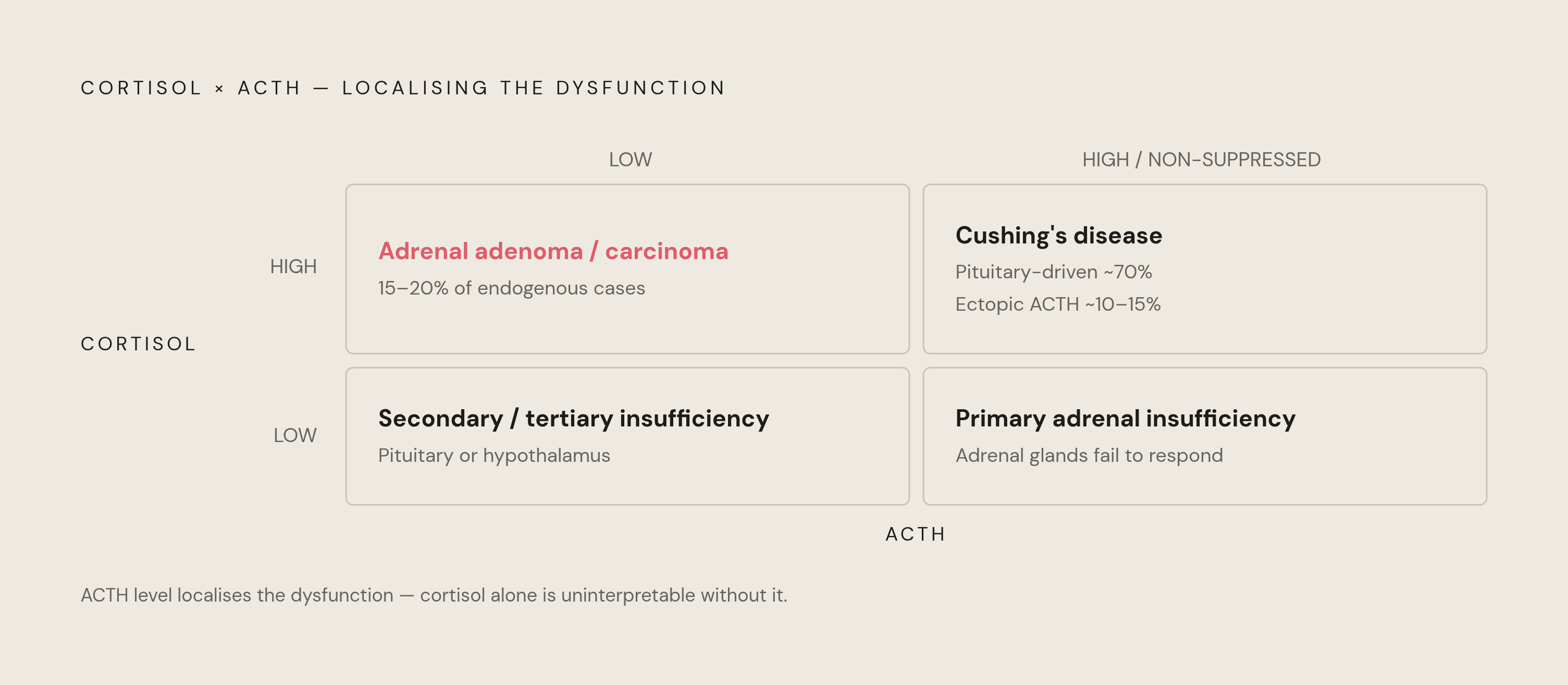
Cortisol tells you what came out the far end of the axis. ACTH tells you where the problem actually sits inside it, and treating the two as interchangeable is a mistake that costs real diagnostic accuracy. Under normal feedback, ACTH rises when cortisol runs low and falls when cortisol runs high, the loop working exactly as designed. Pairing the two values lets a clinician localize dysfunction instead of just flagging that something's off.
Low cortisol alongside high ACTH points to primary adrenal insufficiency: the pituitary sends the signal correctly, but the adrenal glands don't answer it. Low cortisol alongside low ACTH points further upstream, to secondary (pituitary) or tertiary (hypothalamic) insufficiency, where the signal itself never gets generated. On the high-cortisol side, high cortisol with inappropriately high or non-suppressed ACTH suggests pituitary-driven Cushing's disease, which accounts for roughly 70% of endogenous Cushing's cases, with ectopic ACTH production behind another 10 to 15%. High cortisol paired with suppressed ACTH points instead toward an adrenal adenoma or carcinoma, adrenal tumors making up 15 to 20% of endogenous cases.
Chronic psychological stress breaks the feedback loop in a quieter way than any tumor does: ACTH secretion can become dysregulated relative to cortisol output, a pattern that has been discussed in the context of mood and stress disorders. When basal cortisol falls into that indeterminate 100-to-500 nmol/L range, an ACTH stimulation test using Synacthen probes adrenal reserve directly instead of leaning on a static snapshot. It's a dynamic test, watching how the adrenal glands answer a controlled signal, rather than one measurement taken and left to speak for itself.
ACTH comes with a real logistical catch: it degrades fast in blood. Getting a usable result demands careful pre-analytical handling, and that requirement matters a great deal for any executive panel built around remote or concierge phlebotomy rather than a hospital lab a few steps down the hall.
Collection method choices
Blood draws measure total cortisol, bound and free together, and anything that shifts cortisol-binding globulin, oral contraceptives, pregnancy, acute illness, skews the number without reflecting any real change in HPA activity. Blood still earns its place for adrenal insufficiency workups, Synacthen stimulation testing, dexamethasone suppression testing, and critical care assessment of adrenal function. Its limit is the one this piece opened with: a single morning draw catches one instant and says almost nothing about rhythm by itself. Where the clinical question involves localizing the axis, ACTH has to get drawn at the same time as cortisol, never separately, or the comparison means nothing.
Saliva measures the free, biologically active fraction directly, the 5 to 10% of circulating cortisol actually doing biological work. It's non-invasive, and samples can be collected at home and submitted to a lab, which makes multiple timepoints in a single day realistic outside a clinic. An analysis in Clinical Endocrinology found that 83% of patients preferred home salivary collection over a clinic visit, a compliance edge that matters directly for panels built around multiple daily draws. Late-night salivary cortisol is well suited for suspected Cushing's syndrome, since it targets the nadir, the point cortisol should sit lowest, where Cushing's-driven excess shows up most clearly. Saliva is also the only realistic way to build a full four-point diurnal profile or catch the CAR, since repeated blood draws across a single day present a substantial practical barrier outside a clinical setting. An emerging variant, salivary cortisone, may end up a better surrogate for free serum cortisol than salivary cortisol itself, since salivary cortisol converts to cortisone rapidly once in saliva, though it remains an emerging approach rather than standard practice today.
Twenty-four-hour urinary free cortisol captures the roughly 1% of unbound cortisol excreted unchanged, and because it integrates output over a full day, it averages out episodic spikes instead of catching one moment. It stays independent of cortisol-binding globulin levels, which makes it a cleaner read for patients on oral contraceptives or anyone with altered protein levels. It suits confirming persistent cortisol excess and pairs with late-night salivary cortisol in a Cushing's syndrome workup. Its drawback is practical rather than biological: a complete 24-hour collection depends entirely on the patient actually doing it right, and it measures total load rather than rhythm.
Hair cortisol concentration takes the long view, offering a retrospective window on cumulative cortisol exposure over weeks or months, filling a gap none of the single-timepoint methods touch. It's also the method most likely to get oversold, and the data don't back the claims made for it. A 2026 University of Michigan study in Psychoneuroendocrinology (n=117) tested whether hair cortisol actually reflects measurable HPA regulatory biology in healthy people, and the best statistical model explained just 23% of the variance in hair cortisol concentration, built from feedback sensitivity (8.7%), adrenal sensitivity (8%), and stress reactivity (5.3%), while hair washing frequency alone accounted for another 19%, more than all three biological factors combined. Hair cortisol concentration is not a clean way to quantify stress exposure or stress sensitivity, and the associations that do exist still need testing in independent samples before anyone draws clinical conclusions from them. A related composite, the hair cortisol-to-DHEA ratio, got reviewed in a 2025 systematic review and meta-analysis (PROSPERO CRD420251003364) as a possible marker of chronic stress, and the review found the evidence too thin to call it a validated resilience biomarker at this point.
A question of assay technology underlies all four collection methods, rarely comes up in conversation, and decides whether any of these numbers can be trusted. Automated immunoassays remain the most widely used option, but they lack specificity and swing noticeably from one assay run to the next. LC-MS/MS, liquid chromatography-tandem mass spectrometry, offers a sharper alternative, and which assay technology sits behind a given test shapes the answer just as much as which collection method produced the sample.
Sources
- Frontiers | Chronic stress, cortisol dysregulation, and neurodegenerative vulnerability: mechanistic pathways linking HPA-axis dysfunction to Alzheimer’s disease risk
- Does hair cortisol concentration reflect measurable regulatory biology of the HPA axis in healthy humans? - PubMed
- An Integrative Approach to HPA Axis Dysfunction: From Recognition to Recovery
- Cortisol: Normal Range, High & Low Levels Explained (2026)
- An Integrative Approach to HPA Axis Dysfunction: From Recognition to Recovery - The American Journal of Medicine
- researchgate.net
- Stress Biomarkers in Young Adult University Students Before, During, and After the COVID-19 Pandemic: A Longitudinal Cohort Study