Inflammatory Biomarker Panels Beyond High-Sensitivity CRP
Combining CRP with IL-6 and immune markers reveals which inflammatory pathways actually drive risk.

High-sensitivity CRP tells clinicians that inflammation is present. It does not tell them why, how much danger it signals, or whether it will respond to treatment. That gap is why a growing body of cardiovascular and inflammatory disease research now points toward multi-marker panels, combinations of CRP with IL-6, cell-count ratios, and newer structural markers that together sketch a fuller picture of risk than any single number can.
The scale of the problem starts with how common an elevated hs-CRP reading actually is. Analysis of a national health survey dataset found that 52% of adults adults carry hs-CRP levels at or above 2 mg/L, the threshold most guidelines treat as a risk-enhancing factor. That is a coin-flip result across the general population rather than a rare finding flagged in a handful of high-risk patients. It is a coin-flip result across the general population, which means half of the adults walking into a primary care visit already carry a marker that current ACC guidance factors into their cardiovascular risk calculation. A scientific statement from a national cardiology organization found that in patients with established atherosclerotic disease, hs-CRP predicts future cardiovascular events and death at least as well as a standard cholesterol marker.
The problem is what hs-CRP actually measures. It is a downstream acute-phase reactant rather than a signal the body generates directly in response to vascular injury. It is a downstream acute-phase reactant, synthesized by the liver only after interleukin-6 has already done its work upstream. An elevated CRP signals that something is wrong, but says nothing about which process is causing it, how severe it is, or whether a given treatment will actually help. CRP plays no causal role in cardiovascular disease itself, and its levels rise in response to a wide range of conditions beyond vascular injury. An elevated reading can reflect obesity or an unrelated infection just as much as coronary plaque instability, so it is not specific.
The ACC's September 2025 statement, led by writing committee chair George A. Mensah, MD, calls the evidence behind hs-CRP screening "no longer exploratory but compelling and clinically actionable," and it recommends broader screening, particularly for patients in secondary prevention. Yet that same statement acknowledges directly that other biomarkers carry predictive value of their own. And the field's confidence took a real hit in late 2024, when the CLEAR SYNERGY (OASIS 9) trial found that colchicine, an anti-inflammatory drug, failed to reduce major adverse cardiac events in post-MI patients after PCI. Lowering inflammation, in other words, does not automatically buy a patient anything. That result alone should make clear why a single downstream marker, however well-validated, can't tell a clinician which inflammatory pathway is active, how severe it is, or whether it's even treatable. That's the gap a panel is built to close.
What a multi-marker panel adds that a single biomarker cannot
Systemic inflammation doesn't run on one channel. Innate immune activation, the acute-phase response, platelet-neutrophil signaling, and fibrotic remodeling are distinct biological processes, and each leaves a different fingerprint depending on which marker class is doing the measuring. CRP, IL-6, and fibrinogen are among the most studied inflammatory markers for characterizing patients with atherosclerosis, diabetes, and heart failure, three conditions increasingly understood as inflammatory disorders in their own right rather than purely metabolic or structural ones.
The logic of a panel follows the cascade itself. IL-6 sits upstream as the causal driver. CRP and fibrinogen sit downstream as acute-phase responders. Cell-count ratios like the neutrophil-to-lymphocyte ratio capture a different axis entirely, the balance of innate versus adaptive immune activity. Tissue-specific markers like galectin-3 or GDF-15 pick up structural remodeling that none of the above can see. Stacked together, these markers cover different moments in a process that no single test can capture on its own.
Cost matters here too, and it cuts in the panel's favor. Some additions, like the CBC-derived ratios, cost nothing beyond a routine blood draw already being run. Others, like IL-6 assays or galectin-3, require dedicated testing that adds real expense. That means panel composition can flex with the clinical setting and the budget available, rather than demanding an all-or-nothing commitment. The ACC's 2025 statement is candid about where this land: alternative biomarkers "have also found predictive value but generally have not proven superior to hsCRP and often are not widely available in standardized commercial formats." Panels add context, they don't replace the workhorse test.
What a panel does that hs-CRP alone cannot is find the patients CRP misses entirely: someone with a normal CRP but an elevated IL-6 or NLR, a patient whose inflammation is bacterial rather than viral (a distinction procalcitonin can make and CRP cannot), or a heart failure patient whose fibrotic remodeling risk raises galectin-3 well before it raises a natriuretic peptide.
IL-6 as the upstream node: what measuring the driver rather than the signal reveals
CRP exists because the liver responds to IL-6. That single fact reorders the whole conversation: IL-6 is the cause, CRP is the echo, and measuring the echo will always lose some information the original signal carried. Research using causal inference methods supports a more direct mechanistic role for IL-6 signaling in cardiovascular disease than has been demonstrated for hs-CRP. hs-CRP has not cleared that bar. It correlates. It does not cause.
The longitudinal data backs this up repeatedly. A Framingham Heart Study substudy found elevated IL-6 associated with a 68% increased risk of developing chronic heart failure, versus a 2.8-fold increased risk tied to serum CRP at 5 mg/dL or higher, numbers measured on different scales but pointing the same direction. The Health-ABC study, which followed 2,610 elderly adults with no prior heart failure for a median of 9.4 years, found IL-6 associated with a 29% increased risk of developing HF against a 9% increased risk tied to CRP. More telling: across these studies, IL-6 consistently emerged as the stronger independent predictor, with CRP showing comparatively weaker associations once other inflammatory markers were accounted for. The MESA study, covering 6,622 apparently healthy adults, found elevated IL-6 strongly predictive of mortality, adverse cardiovascular outcomes, and incident heart failure. A 2021 case-cohort study extended the pattern to heart failure with preserved ejection fraction, finding a significant association between IL-6 and incident HFpEF in the general population.
The therapeutic story complicates things in an instructive way. Ziltivekimab, a monoclonal antibody that targets the IL-6 ligand directly, was evaluated in patients with high atherosclerotic risk and shown to reduce inflammatory biomarker levels in early-phase studies. That set up larger outcome trials, including ZEUS in patients with established ASCVD, CKD, and systemic inflammation. ZEUS reported that the drug lowered free IL-6 and hs-CRP exactly as designed. It did not reduce major adverse cardiac events. That result is the central epistemic problem panels have to sit with: a biomarker can fall in response to treatment without the patient getting any safer. It's also, paradoxically, the argument for tracking more than one marker at once, since no single number, however well it moves on a drug, guarantees the outcome that matters.
Part of why IL-6 carries more mechanistic weight than CRP traces back to where it's made. Macrophages, monocytes, endothelial cells, vascular smooth muscle cells, and fibroblasts all produce it, reflecting activity happening directly at the vessel wall and in the tissue itself, not just a hepatic response manufactured several steps downstream.
CBC-derived inflammatory ratios: the cost-free extension of any panel
The neutrophil-to-lymphocyte ratio and the platelet-to-lymphocyte ratio come from numbers already sitting in a routine complete blood count. No extra vial, no extra assay, no extra line item. That alone makes them the easiest expansion of any inflammatory panel a clinician could order.
NLR captures the tension between inflammatory drive, reflected in neutrophil counts, and adaptive immune response, reflected in lymphocyte counts. Some researchers argue the ratio outperforms either cell count on its own for predicting cardiovascular conditions ranging from hypertension and heart failure to infective endocarditis, stable coronary disease, and acute coronary syndrome. A meta-analysis pooling 13 high-quality prospective cohorts run between 2019 and 2025 looked at IL-6, hs-CRP, and NLR together across diverse geographic regions and clinical settings. All three markers stayed independently associated with mortality and adverse heart failure outcomes across diverse geographic regions and clinical settings. That's a fairly demanding statistical bar to clear, and NLR cleared it using data most labs already have on file.
PLR adds a thrombotic dimension on top of the inflammatory one, which makes it particularly useful in settings where troponin-based scoring isn't practical or available. A more recent addition, the mean platelet volume-to-lymphocyte ratio, was studied in 103 elderly heart failure patients admitted to Suzhou Hospital (Anhui Medical University), evaluating NLR, PLR, and MPVLR together in elderly heart failure patients. MPVLR layers a platelet-activation signal onto the same lymphocyte denominator the other ratios use.
None of this is settled science yet. Heterogeneity in study design, in when blood was drawn relative to symptom onset, in assay standardization across labs, and in the populations studied has kept these ratios in promising-but-not-proven territory rather than earning them a spot in formal guidelines. Still, in any setting where a CBC is already being drawn, which is nearly everywhere, adding NLR and PLR to the read costs nothing and adds a real signal. The bar for including them in practice is about as low as it gets.
Novel acute-phase reactants, SAA, procalcitonin, presepsin, and calprotectin, and where they earn their place
These markers earn their keep in a different arena than the cardiometabolic space discussed above. Their strongest evidence sits in acute settings, sepsis, pneumonia, active bacterial infection, and treating them as chronic risk-stratification tools would stretch the data past where it actually holds.
Serum amyloid A moves in a range CRP can't match, rising 100- to 1,000-fold over a baseline of roughly 1 microgram per milliliter during an acute-phase response. A 2025 study of community-acquired pneumonia patients found SAA levels at emergency department admission markedly higher in non-survivors than survivors, with an AUC of 0.768 for predicting mortality, outperforming both procalcitonin (0.671) and hs-CRP (0.657) in that dataset. The optimal SAA cutoff identified was 280.2 mg/L, with 79.4% sensitivity and 68.5% specificity. But a separate 193-patient comparative study found a different ranking when sepsis specifically was the target: AUCs of 82.6% for SAA, 82.0% for CRP, 90.3% for procalcitonin, and 85.7% for presepsin. Procalcitonin and presepsin came out ahead there. No single marker dominates across every acute inflammatory context, and that inconsistency itself is the finding.
Procalcitonin's real advantage is specificity: it rises sharply in severe bacterial infection, offering a degree of pathogen-type discrimination that broader acute-phase reactants do not provide. Calprotectin, a neutrophil-derived protein, has been studied as an early marker in the course of sepsis and bacterial infection. Presepsin, in that same 193-patient study, posted the second-best AUC behind procalcitonin at 85.7%.
The pattern across all four points toward combination rather than substitution. Pairing SAA with procalcitonin, CRP, and presepsin appears to sharpen diagnostic accuracy for early sepsis beyond what any one marker delivers alone, improving specificity without giving up sensitivity. In chronic cardiometabolic risk work, this whole category adds little. Its value lives in acute triage and infection characterization, and choosing which of these tests to run should follow directly from what the clinician actually needs to know.
Emerging structural and fibrotic markers: galectin-3, pentraxin-3, and GDF-15
Galectin-3 measures something CRP and IL-6 simply don't touch: structural remodeling. The protein is implicated in macrophage activation and myocardial fibrosis, mechanisms distinct from the systemic inflammatory signaling the earlier markers reflect. Research consistently flags Gal-3 as a leading candidate biomarker for cardiovascular disease, with a role that spans tracking disease progression to forecasting outcomes. It also complements existing heart failure diagnostics rather than competing with them, adding signal on top of natriuretic peptide testing rather than duplicating it. A 2025 proof-of-concept study involving 131 patients undergoing coronary angiography found Gal-3 values more than doubled in patients with significant chronic coronary syndrome compared to controls, a result pointing at structural disease severity rather than generic systemic inflammation.
Pentraxin-3 offers a different kind of specificity. Unlike CRP, which the liver manufactures as a hepatic acute-phase protein, PTX3 gets produced locally, right at the site of vascular inflammation. That local origin may make it a sharper indicator of what's actually happening in the vessel wall, rather than a systemic average diluted by whatever else is going on in the body.
GDF-15 tracks yet another axis: oxidative stress and mitochondrial dysfunction across a range of cell types, cardiomyocytes included. None of the cytokine or acute-phase markers discussed so far touch that biology at all.
All three remain emerging rather than guideline-endorsed. The evidence base is growing but uneven across studies, and their strongest current use case is as a prognostic add-on in heart failure and atherosclerosis rather than a frontline screening tool. How these markers interact with established risk factors, dyslipidemia, insulin resistance, elevated NT-proBNP, is still being worked out. That unresolved complexity is itself a reason to think in terms of structured panels rather than ordering tests one at a time as they come to mind.
How to think about panel composition in clinical practice
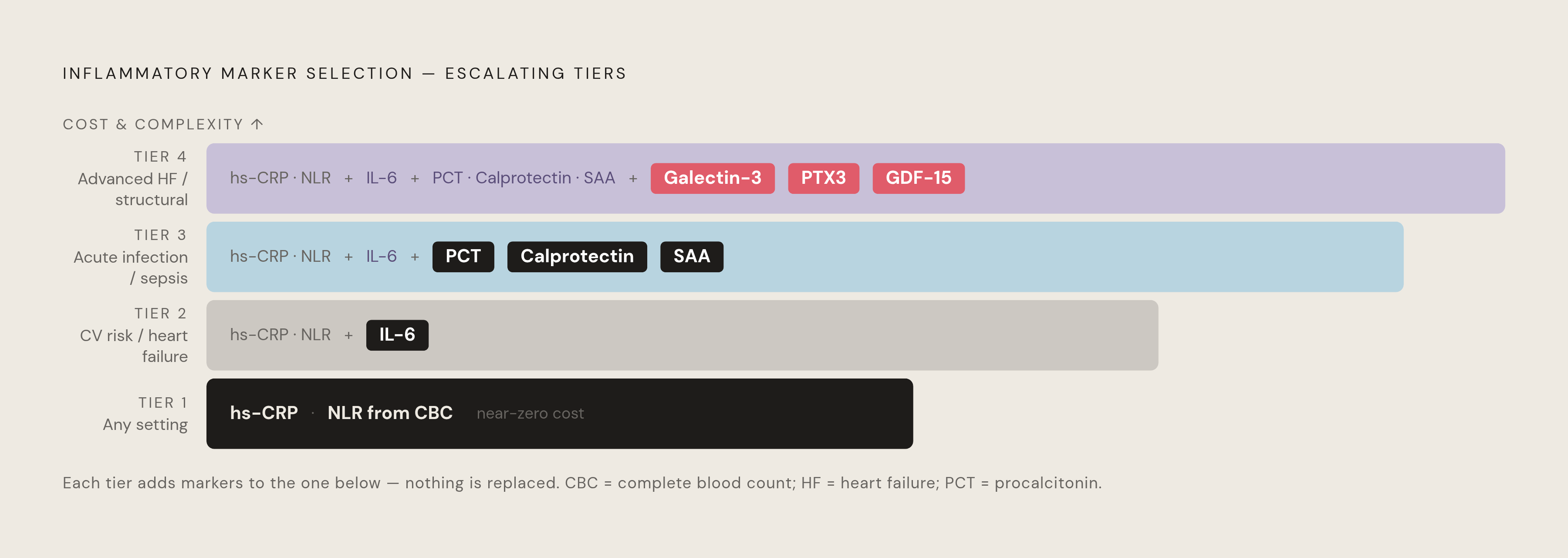
There's no single panel that fits every patient, and pretending otherwise misreads the whole point of layering markers in the first place. Chronic cardiometabolic risk stratification, acute infection triage, heart failure prognosis, and treatment-response monitoring are four different clinical questions, and each calls for a different set of tests.
A tiered approach tracks the evidence reasonably well. Tier one, appropriate in essentially any setting given how little it costs: hs-CRP alongside NLR pulled from a CBC already being drawn, giving a baseline inflammatory signal plus a cellular ratio at no marginal expense. Tier two, for cardiovascular risk and heart failure specifically: add IL-6, since it captures the upstream causal signal Mendelian randomization studies support, and since it held its predictive value in multivariable models where CRP dropped out. Tier three, for acute infection or sepsis triage: add procalcitonin and calprotectin for bacterial specificity, and SAA where the assay is available, given its mortality-stratification performance in pneumonia. Tier four, reserved for advanced heart failure, structural disease, or research-adjacent practice: galectin-3 for fibrotic remodeling, PTX3 for local vascular inflammation, GDF-15 for oxidative and mitochondrial stress.
These markers aren't static numbers frozen at diagnosis. The ACC's guidance notes hs-CRP responds to behavioral change: smoking cessation drops it by roughly 0.40 mg/L in patients with established cardiovascular disease, aerobic exercise by 0.59 mg/L in healthy adults and 0.34 mg/L in those with existing disease, and a Mediterranean diet by 0.98 mg/L per a meta-analysis of randomized trials. Tracked over time, panel markers show whether behavioral and pharmacologic interventions are actually doing something, not just whether a diagnosis has been made.
The ZEUS trial result produces the honest caveat that qualifies all of this, and its data stand as direct evidence of that limit. Ziltivekimab lowered both IL-6 and hs-CRP exactly as intended, and MACE didn't budge. Panels inform risk, and they track biology over time, but the therapeutic meaning of any single elevated marker still needs to be read in the context of the whole clinical picture, not acted on as if the number alone were the diagnosis.
Sources
- Inflammatory Markers - American Society for Preventive Cardiology
- Systemic Inflammatory Biomarkers (Interleukin-6, High-Sensitivity C-Reactive Protein, and Neutrophil-to-Lymphocyte Ratio) and Prognosis in Heart Failure: A Meta-Analysis of Prospective Cohort Studies - PMC
- Inflammatory Biomarkers in Heart Failure: Clinical Perspectives on hsCRP, IL-6 and Emerging Candidates - PMC
- Inflammation and Cardiovascular Disease: 2025 ACC Scientific Statement: A Report of the American College of Cardiology | JACC
- pmc.ncbi.nlm.nih.gov
- rheumnow.com